Scarring case study - right upper limb and left lower limb
The aim of this case study is to illustrate that an assessment of scarring should provide a clear description of the scar/s that in turn supports an assessment with reference to all 5 of the TEMSKI criteria and all 10 of the TEMSKI descriptors. Reasoning related to the assessment of multiple scarring must also be included.
Motor Accident Details
39 year old driver of a small car which collided with a four-wheel drive. Assessment 2 years post motor accident. Clinical appearance of mature scars. No further treatment planned.
Injuries:
- Laceration right forearm
- Closed comminuted fracture left patella
- Scattered lacerations
Surgery:
- Debridement and repair laceration right arm and left hand
- Repair partially divided tendon left extensor carpi ulnaris tendon
- Open reduction and internal fixation left fractured patella
- Subsequent removal of internal fixation from his left knee.


Description:
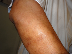
- Right arm: Fine, soft, flat, mobile scar, 10 cms long x 3 cms wide, on the posterior aspect of the proximal end of right forearm; no stitch marks; very minor contour deformity; small muscle hernia visible and slightly raised above the level of surrounding skin beneath the scar. No effect on ADLs; no treatment required.
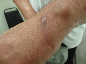
- Left hand: small scar, 5 cms long, ulnar side of the dorsum of left hand; majority of scar fine, radial end of scar widened and measured 1.0cms x 0.8 cms; scar soft and mobile; slightly hyperpigmented; no stitch marks; no contour deformity. No effect on ADLs; no treatment required.
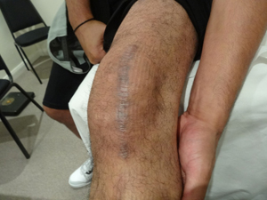
- Left Knee: prominent, longitudinal scar, 15 cms long and up to 1.4 cms wide over patella of left knee; scar soft, flat and mobile; hyperpigmented; no stitch marks; no contour deformity. Unable to tolerate prolonged kneeling which impacted ADLs in relation to cleaning and gardening; no treatment is required.
Assessment:
- Conscious of the scarring;
- Noticeable colour contrast of the scarring with surrounding skin;
- Easily able to locate scars;
- Minimal trophic changes:
- Stitch marks barely visible:
- Anatomical location visible:
- Very minor contour defect as a result of the small muscle hernia:
- Minor limitation of a few ADLs in relation to the left knee:
- No treatment required; &
- No adherence.
Determination:
Overall the 3 scars best fit is in the 1% - 2% WPI range.
Reasoning:
Several factors warrant the allocation of 2% WPI. The total effect of the scars on the organ system as a whole has taken account of: the multiplicity of the scars; the visibility of all 3 scars; the minor contour defect; and, there is a minor limitation in the performance of some ADLs.
